Cardiologist’s specialized technique brings relief to patients – young and old.
By Steve Sullivan
Though decades apart in age, Zane Williams and Merle VanGorpen share the same serious heart condition.
They both have Wolff-Parkinson-White (WPW) syndrome, and both have successfully received treatment for it at Mary Greeley Medical Center.
WPW is the result of an abnormal electrical pathway between the upper and lower levels of the heart. This pathway can, in a sense, reroute electrical signals in the heart and send them in directions they should not go. It’s a form of arrhythmia and can cause a very rapid racing heart rhythm called “supraventricular tachycardia.” This pathway can also be associated with lethally rapid transmission of atrial fibrillation, which can result in ventricular fibrillation—otherwise known as cardiac arrest. It is typically diagnosed through an electrocardiogram (EKG), which charts the heart’s electrical activity and indicates problems.
People are born with WPW and symptoms can arise at any age, or sometimes never. While some symptoms can be treated with medicine, a procedure called cardiac ablation
is often used to correct WPW.
Ablation is a minimally invasive procedure that is aimed at killing the tissue causing the arrhythmia. In the case of WPW, killing the bad tissue can block that abnormal pathway and help ensure that electrical signals are going in the right directions.
Dr. Denise Sorrentino, an electrophysiologist and cardiologist with the Iowa Heart Center in Ames, performed ablations on Williams and VanGorpen. She is part of a team of cardiologists from Iowa Heart Center and McFarland Clinic that provide cardiac care at Mary Greeley for patients from throughout central Iowa. Sorrentino has treated patients and been on call at Mary Greeley for the past 21 years.
“Ames has always been my home base,” she said. “A lot of my patients were traveling, as was I, and Mary Greeley was willing to purchase new advanced mapping equipment for ablations. I’m happy to be bringing
it back home.”
So, obviously, are Williams and VanGorpen.
The Retiree
VanGorpen is a retired insurance claims adjuster from Webster City who enjoys camping trips with his wife.
A few years ago he woke up in the middle of the night and “could tell my heart was skipping a beat.” He sought medical attention and was diagnosed with WPW.
He didn’t have any problems until last September. During a camping trip near Colo, he passed out while walking to the campground bathroom. He didn’t get too worried about that incident. A week later, though, he had to take the camper to a dealership for repair.
“I was leaving the dealership, driving my car out of the lot and the next thing I knew I ran into a fence across the road,” he said. “I had no sensation or anything. I just lost consciousness.”
He sought medical attention again, and the thinking was that he might have had an adverse reaction to some sinus medicine. A few days later, he was home watching television when he started sweating and feeling a tightness in his chest. He ended up at Mary Greeley for seven days while his heart was monitored. Ultimately, he had an ablation.
The Athlete
Williams is a junior at Webster City High School, where he’s on the football and wrestling teams. At the start of the 2017 football season, Williams would come home from practice with his heart racing.
“It was beating really fast and it worried me but I waited a few weeks before saying anything. I didn’t want to overreact,” he said.
On Sept. 22, in a victory over Ballard High School, Williams, a tailback, rushed for 179 yards and scored four touchdowns. Football was going great, but he knew something wasn’t right. Urged by his older sister, Williams finally talked to his parents about what he was experiencing. They got him to a doctor for tests. On Sept. 27, Williams was diagnosed with WPW, putting his high school football career on an extended time-out.
“I was down in the dumps after getting the news. I was in my prime, playing football,” he said. “I had good people around me, my family and teammates. A good support system. They told me I would only miss three games and would be back at it.”
WPW can be scary regardless of someone’s age, but it can be particularly tough for young people to face.
“A lot of my patients are younger, and they often find themselves in the waiting room, sitting beside people much older than them, and thinking that something must be terribly wrong,” Sorrentino said.
The stress of a diagnosis like this can “take a big toll on patients and parents. I spent a lot of time with young patients and their families,’ she said. “I work to assure patients of all ages that arrhythmia is something we see in otherwise healthy people and ablation is 98 percent curative.”
Williams had his ablation on Oct. 11.
Tissue Burn
Williams and VanGorpen both had radio frequency (RF) ablations performed by Sorrentino.
An ablation patient is usually under conscious sedation, which induces a medium to deep sleep. General anesthesia usually isn’t necessary.
To perform the ablation, Sorrentino inserts thin, flexible wires, or catheters, through arteries in the groin and threads them to chambers in the heart. Typically there will be 2-3 catheters on either side of the patient’s body. Each has a purpose.
The patient will have patches placed on the chest to read EKG signals sent from one of the catheters. These signals will be used to create a 3D map, which will guide Sorrentino to the place where the ablation needs to be done.
Other catheters are strategically placed to measure electrical conduction in the heart. Signals can be sent through the wires that will intentionally jump start an arrhythmia. This aids in the mapping of the tissue where the arrhythmia is originating.
“A lot of data and experience goes into this procedure,” said Sorrentino. Based on the location of the arrhythmia, Sorrentino will perform either a RF ablation, which uses heat to kill the bad tissue, or cryoablation, which kills the tissue through freezing.
The arrhythmia may be close to the heart’s normal electrical system, an area known as the AV node. This area is a sort of electrical relay station between the upper and lower chambers of the heart. If this is the case, Sorrentino will likely kill the bad tissue with an -85° Celsius (-121° Fahrenheit) cryoablation blast. This method is less likely to impact tissue surrounding the AV node.
If the problem is farther from the electrical system, then RF is used. Following the 3D mapping, Sorrentino maneuvers an irrigated tip that delivers a stream of saline while the catheter is burning bad tissue. The saline helps control the burn and support cooling, allowing Sorrentino to go deeper into tissue, ensuring she is permanently fixing the source of the arrhythmia.
“Cryoablation is painless. The patient won’t feel anything. We tell a patient going through radio frequency ablation that they might feel a burning sensation during the procedure,” she said.

Watch an Ablation
Learn more about ablation, and see Dr. Denise Sorrentino perform the procedure:
Aftermath
VanGorpen is doing fine now and looking forward to nice weather for more camping trips. He’s watching his diet, cutting down on salts and fats, and getting in some exercise whenever he can. After years of excellent health, he jokes that he finally has some aches and pains to share with pals who always seem to have an ailment to talk about.
“You come to the realization that you’re no longer 29 years old and can do whatever you want,” he said.
Nine days after his ablation, Williams suited up with his team and returned to the gridiron.
“I was nervous at first, but once I got that first snap in, I did what I do,” he said.
Williams ran for 145 yards and scored two touchdowns, helping Webster City defeat Boone High School and earn a spot in the district playoffs. That and his successful ablation were not all he had to celebrate. During his break from football, Williams was elected Webster City High’s homecoming king. After football, he moved onto wrestling, achieving his 100th career victory this season.
Picture Your Heart

Mary Greeley’s new cardiac CT service
offers many benefits for patients.
Mary Greeley Medical Center has expanded its cardiac care services with the addition of advanced cardiac CTA technology.
Many of the unit’s patients initially receive treatment at Mary Greeley. However, the unit is increasingly receiving referrals for patients who have had treatment in Des Moines, Iowa City and other locations outside of Ames.
A cardiac CTA is a non-invasive procedure that provides highly detailed images of the heart and the coronary arteries. The images can indicate whether the arteries have calcium buildup or blockage, and help providers determine a course of treatment.
Here’s what you need to know about this new service. Special thanks to Scott Cue, Mary Greeley’s director of Radiology Services, McFarland Clinic cardiologist Dr. Jason Rasmussen, and McFarland Clinic radiologist Dr. Doug Lake, for their expert info on cardiac CTA.
What is a cardiac CTA?
CTA stands for computed tomography angiography. A CT scan is a medical test that produces multiple images of the inside of the body. It is often used for scans of the brain. The images provide much greater detail than a traditional x-ray, particularly of soft tissues and blood vessels.
Angiography is a medical imaging technique used to visualize the inside of the blood vessels.
Cardiac, of course, refers to the heart so a cardiac CTA is a medical test that generates a detailed image of that particular organ.
Why does my doctor want me to have a cardiac CTA?
Your doctor may want you to have a cardiac CTA for any number of reasons. You most likely have been experiencing symptoms that could be indicative of heart disease. You could have had a cardiac stress test that raised questions,
or you may have some of the major risk factors beyond family history and that combined with your symptoms have raised a red flag. Those risk factors include high cholesterol, diabetes, high blood pressure, smoking, and being overweight and/or physically inactive.
How is a cardiac CTA performed?
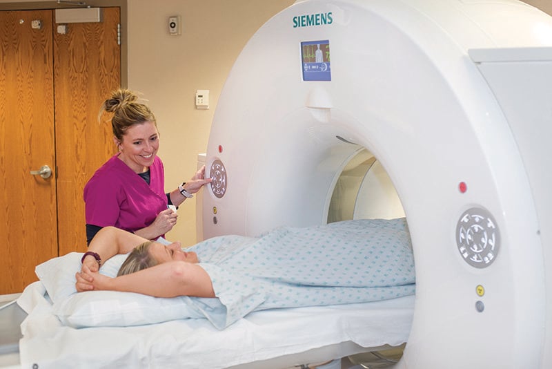
A CT technologist will help you lay flat on your back on the exam table. Electrodes will be attached to your chest so that the electrical activity of your heart can be monitored during the procedure.
The CT scanner rotates once every 0.28 seconds, collecting a multitude of photos encircling the patient in a very short amount of time. This speed is beneficial to the comfort of the patient.
A slow and regular heart rate allows the best image quality, so you will likely receive a medication to slow your heart rate and make it more regular.
How long does a cardiac CTA take?
From the time a patient walks into the CT room to the time they walk about, it usually takes anywhere from 30 to 60 minutes. Patients usually arrive 90 minutes before the exam for a full nursing assessment and to ensure the patient’s heart rate and rhythm are ideal.
What is the difference between an angiogram and a cardiac CTA?
An angiogram, which also captures pictures of the heart, is an invasive procedure. In that procedure, a tube called a catheter is placed into a blood vessel in either the groin or the arm and is guided to the heart. With a cardiac CTA, a catheter does not have to be placed.
A patient will have an IV during the procedure to administer a contrast dye. That allows for better, more enhanced pictures of the heart to be taken and helps to differentiate the blood vessels from the rest of the heart muscle.
What are the benefits to having a cardiac CTA done over an angiogram?
Since the cardiac CT is a non-invasive procedure, no sedative is required. That means that once the procedure is over a patient is able to walk out and resume normal activities immediately. It takes very little time and is painless.
Also, scans from a cardiac CTA are read by either a radiologist or a cardiologist. That means a patient gets a highly trained physician to interpret findings and work with the patient care team to determine the best course of action.
Your cardiologist will determine whether an angiogram or a cardiac CTA is the best option for you.